Unveiling Heart Health: 5 Key Abnormalities Found on an Echocardiogram
Heart disease is a leading cause of death in Americans, and it is increasing at an alarming rate. It affects how our bodies supply essential nutrients and oxygen to organs. It creeps in with no signs and silently, reshaping our daily mood and mobility. Many elements contribute to its impact, such as poor diet, inactivity, and stress, and making critical lifestyle changes can be a line of defence. Understanding the 5 abnormalities on the echocardiogram can help you spot the troubles early.
What is an Echocardiogram?
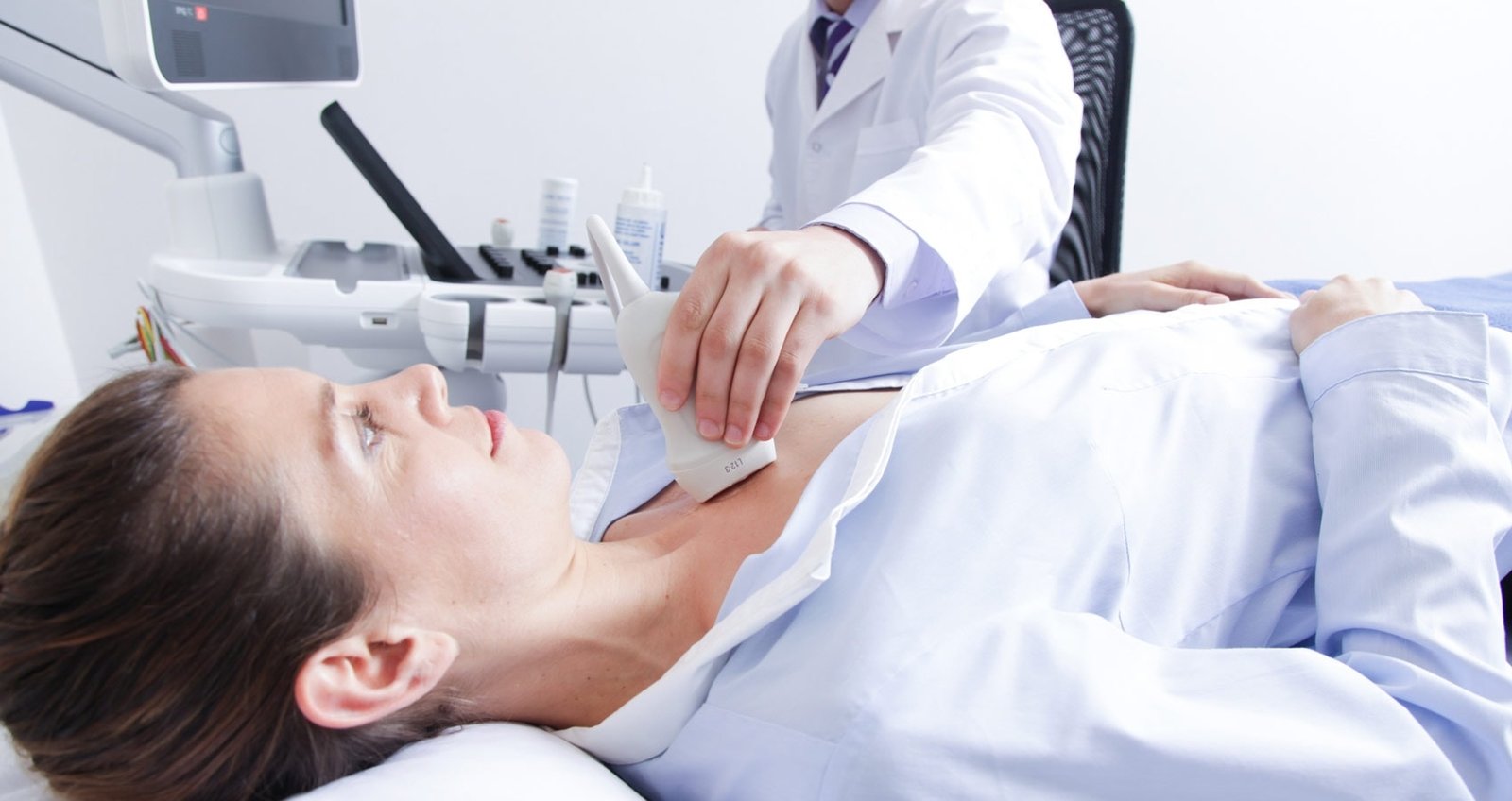
An echocardiogram or cardiac ultrasound is a special test that uses sound waves to produce detailed and live pictures of the heart. It is a radiation-free test and a noninvasive exam in which a trained sonographer swabs an ultrasound wand across the chest, delivering sound waves that bounce back within the wand. This information is sent to the computer, creating two-dimensional or three-dimensional images of the beating heart, valves, and other structures.
In America, heart disease is a leading cause of death in most adults, with approximately 695000 deaths yearly. Coronary heart disease remains the most common type of heart disease, killing 371,506 people in 2022. About 1 in 20 adults over age 20 has coronary artery disease (CAD), and echocardiography has proven to be a valuable tool for diagnosing and managing the condition.
Normal Vs. Abnormal Echocardiogram
Recognizing regular vs. abnormal echocardiogram results helps take proactive steps to preserve the functions of the heart.
Normal Echocardiogram
- The ejection fraction is within 55%-70%
- The pericardial space is free of excessive liquid
- The valve motion is smooth with no stenosis or regurgitation
Abnormal Echocardiogram
- Wall motion abnormalities, such as akinesis, that suggest infarction
- Enlarged chambers that dictate pressure overload
- Pericardial effusion is visible as fluid around the heart
5 Key Abnormalities Found on an Echocardiogram
1. Valve Abnormalities
The valves are one-way passages that ensure blood flows through the chambers. When the valves malfunction, either by leaking or narrowing, the heart works harder to compensate, often causing symptoms initially. In such a case, an echocardiogram is an excellent option for spotting such issues early.
- Color Doppler highlights the turbulence, answering what colors are bad on an echocardiogram
- Stenosis– the valve opening is too tight, indicating tricuspid, mitral, aortic, and pulmonary conditions
If not treated well, valve disease would ultimately lead to heart failure. However, early detection of such abnormalities at the Healwell Primary Clinic can lead to tailored treatment for abnormal echocardiogram findings before severe symptoms occur.
2. Chamber Size and Function Abnormalities
Your heart chambers need the right strength and size to pump blood efficiently. Enlarged or shrunk chambers signal underlying strain or damage affecting overall blood flow. An echocardiogram measures the dimensions and ejection fraction of the blood, a key marker of heart performance.
- Low ejection fraction echo– shows the decreased pumping capabilities of the heart
- Cardiomegaly– expanded ventricles or atria, indicating pressure overload
- Diastolic dysfunction- impaired relaxation can be seen in how an echocardiogram is done on a woman with diabetes or hypertension
At Healwell Primary Care, we make a close differentiation between normal and abnormal echocardiogram results to create personalized plans, helping you restore functionality and vitality.
3. Wall Motion Abnormalities
The heart’s muscle walls contract to circulate blood. Pronounced motion defects in an area can reveal prior damage from a heart attack. An echocardiogram is helpful in this case to visualize these motion patterns in real time, helping clinicians make precise interventions.
- Hypokinesis– a decreased movement in one segment
- Dyskinesis- paradoxical bulging during contraction
- Akiensis– A complete lack of movement
Identifying heart wall motion abnormalities early prevents further muscle loss. Our team at Healwell Primary Care uses the test results to recommend angioplasty, stress tests, and different rehab programs that strengthen the heart.
4. Myocarditis & Pericardial Effusion
A fluid-filled sac surrounds your heart, called the pericardium, that can become overwhelmed by inflammation or excess fluid. Whether post-viral or idiopathic, such conditions can exert pressure on the heart, impacting blood flow.
- Effusion– abnormal fluid accumulation around the heart (> 50 mL)
- Tamponade Risk– When fluid compression impairs heart function
The pericardial conditions can become life-threatening if left untreated, and they can make a significant impact on heart health.
5. Intracardiac Thrombus & Strain Abnormalities
Beyond the heart’s structure, echocardiograms help detect blood clots that can lead to various complications, including stroke, pulmonary embolism, and other systemic embolisms. Clots inside the chamber increase the risk of stroke, while strain imaging uncovers early dysfunction before the ejection fraction fails.
- Blood clot in heart echocardiogram– it identifies the thrombus that could embolize
- Chemotherapy cardiotoxicity screening to protect the vulnerable patients
- Global longitudinal strain or GLS flags early cardiomyopathy
An echocardiogram explains the abnormalities, allowing your doctor to intervene sooner and reduce complications.
What should you not do before an echocardiogram?
There are several aspects to notice before going for an echocardiogram test.
- Skip caffeine– avoid caffeine, as it can alter heart rate and Doppler flow pattern.
- Dress comfortably– wear comfortable clothes with easy chest access, as the test will be performed on the chest.
- Take light meals– before a stress echo, avoid heavy meals.
Following these tips can help ensure clean and artifact-free screening.
How is an Echocardiogram Done on a Woman
The procedure for conducting an echo test is similar for both men and women. It’s a simple, painless procedure providing critical insight/details into heart function tests. You won’t need to undress; open your top and lie comfortably. The technician will apply gel to the chest area and glide a probe through the various positions, capturing heart views.
What happens after an abnormal Echocardiogram?
The results of an abnormal echocardiogram open the door to tailored care. At Healwell Primary Care clinic, we translate the findings into a detailed action plan. We help you guide from interpretation to a clear and improved outcome.
- During this session, the cardiologist will discuss your echocardiogram results in detail, explain the implications of any abnormalities, and recommend a personalized plan for further evaluation or treatment.
- Stress echo, CT/MRI, and other procedures are performed
- The decision between medication, lifestyle coaching, and various procedures is assessed for your personal treatment plan
The multidisciplinary team approach at Healwell Primary Care ensures that you understand each aspect of the findings and feel supported at each stage. We provide the reassurance and guidance you need during this process, ensuring you feel cared for and secure in your journey to better heart health.
Personalized treatment at Healwell Primary Care
The silent progression of heart disease underscores the importance of early detection and informed decision-making. Understanding the normal vs. abnormal echocardiogram results will help you find proper care. At Healwell Primary Care, our experts leverage advanced echocardiography equipment to craft a personalised treatment plan that combines lifestyle coaching with cutting-edge interventions. Schedule an appointment now with a cardiologist at Healwell Primary Care to schedule a detailed echocardiogram, and let us guide you toward a healthier lifestyle.
FAQs:
1. What is an echocardiogram?
An echocardiogram is a non-invasive ultrasound test that helps visualize your heart chambers, walls, and valves.
2. How is an echocardiogram done on a woman?
An echocardiogram is identical regardless of gender. A gel is applied to the chest, a transducer is moved over the skin, and images are captured. The process is simple, painless, and completed under 30 minutes.
3. How long does it take to get an echo result?
In most cases, echocardiogram images are interpreted immediately, but you can expect a detailed report within 24 hours.
4. How does Healwell Primary Care handle abnormal echocardiogram results?
The clinic team reviews the test results with you, recommends follow-up tests, and develops individual treatment plans.
5. How do I schedule an echocardiogram test?
You can call our office at (312)971-7147 or visit our website to schedule an appointment.


Contact Us Today
Book an in-person or online visit in seconds. Call (312) 971-7147 or click on Book Appointment.